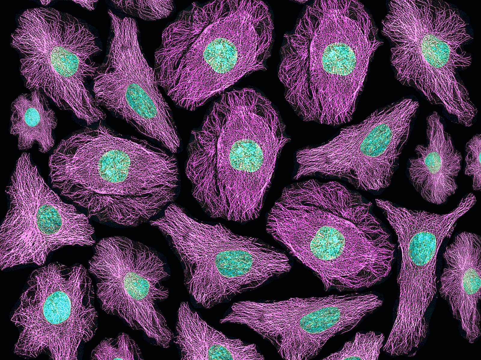
The global market for live cell imaging, a powerful tool for visualizing and analyzing cellular processes in real time, is experiencing robust growth, driven by a confluence of factors that highlight its significance in biomedical research and drug discovery. The market is expected to reach a value of approximately $4.3 billion by 2029, propelled by a compound annual growth rate (CAGR) of 8.4%. This surge in growth can be attributed to the increasing adoption of various screening technologies, particularly in the pharmaceutical industry and academic research centers, where live cell imaging is proving to be a critical tool for understanding the complex interplay of cells in health and disease.
Advancements in Imaging Technologies
The recent advancements in imaging technologies, including the development of high-resolution microscopes and sophisticated fluorescence techniques, have revolutionized the field of live cell imaging. These innovations enable researchers to observe intricate cellular processes in real time with unprecedented precision and accuracy. Moreover, the development of advanced software for image analysis and automation has further enhanced the efficiency and usability of live cell imaging systems, making them indispensable tools in a wide range of biomedical research applications.
High-Resolution Microscopy and Fluorescence Techniques
The ability to capture images at increasingly higher resolutions has significantly advanced our understanding of cellular structures and their intricate functions. High-resolution microscopes allow scientists to visualize cellular components with remarkable detail, providing valuable insights into the complex mechanisms that govern cell behavior. The use of fluorescence techniques, where specific molecules or structures are tagged with fluorescent dyes, has further enhanced the ability to track and analyze cellular processes in real time.
Software for Image Analysis and Automation
The development of sophisticated software for image analysis and automation has revolutionized the way live cell imaging data is processed and interpreted. These software tools enable researchers to quickly and accurately analyze large datasets, identify patterns, and extract meaningful information from the images. Additionally, the automation capabilities of these software solutions have significantly streamlined the live cell imaging workflow, reducing the time and effort required for data acquisition and analysis.
The Rise of Personalized Medicine
The shift towards personalized medicine is a major driver of the live cell imaging market. As medical treatments increasingly focus on tailoring therapies to individual patients, the ability to observe live cellular responses to drugs or treatments in real time is crucial. Live cell imaging enables researchers to study disease mechanisms at the cellular level, leading to more targeted and effective treatments.
Tailoring Treatments to Individual Patients
The concept of personalized medicine relies on the understanding that each individual responds differently to treatments due to variations in their genetic makeup and other biological factors. Live cell imaging plays a crucial role in this paradigm by providing researchers with a powerful tool to observe how individual cells respond to specific therapies. This information is then used to develop customized treatment plans that are tailored to the unique characteristics of each patient, potentially leading to more effective and less invasive therapies.
Drug Discovery and Development
Live cell imaging is an indispensable tool in the drug discovery and development process. It allows researchers to study the effects of new drugs on live cells in real time, providing insights into their potential efficacy and toxicity. By observing how cells respond to different drug candidates, researchers can identify promising compounds that are more likely to be effective in treating specific diseases.
The Impact of High-Content Screening
The rapid adoption of high-content screening (HCS) technologies in drug discovery and development is a significant driver of the live cell imaging market. HCS involves the simultaneous analysis of multiple cellular parameters, such as cell morphology, protein expression, and cell signaling pathways, using automated imaging systems. This high-throughput approach enables researchers to screen large libraries of compounds in a short period, accelerating the drug discovery process.
Accelerating Drug Discovery
The use of HCS in combination with live cell imaging technologies has significantly accelerated the drug discovery process. It enables researchers to identify drug candidates that are more likely to be effective and safe, leading to faster development timelines and reduced costs associated with drug development.
Understanding Cellular Mechanisms
High-content screening provides researchers with a comprehensive view of cellular responses to different stimuli, such as drugs, environmental factors, or genetic modifications. By analyzing these responses, researchers can gain a deeper understanding of the underlying cellular mechanisms involved in disease development and progression. This knowledge is crucial for developing effective therapies and identifying new targets for drug development.
Challenges and Opportunities
While the live cell imaging market is experiencing significant growth, it also faces several challenges, such as the high cost of advanced imaging systems, the complexity of technology and user training, and ethical concerns associated with the use of live cells in research. These challenges present opportunities for innovation and development in areas such as cost-effective imaging solutions, user-friendly software, and ethical guidelines for research.
Cost-Effective Imaging Solutions
The high cost of advanced live cell imaging systems remains a barrier to entry for smaller research institutions and laboratories. The development of more affordable and accessible imaging solutions, such as portable microscopes and open-source software, could broaden the adoption of live cell imaging technologies, allowing a wider range of researchers to benefit from its capabilities.
User-Friendly Software
The complexity of live cell imaging technologies can pose a challenge for researchers who lack specialized training. Developing user-friendly software and intuitive interfaces could make live cell imaging systems more accessible to a broader audience, reducing the barriers to entry for researchers who may not have extensive technical expertise.
Ethical Guidelines
Live cell imaging often involves the use of live organisms, cells, and tissues, which can raise ethical concerns, particularly in stem cell research or genetic modification studies. Developing clear ethical guidelines and regulations for the use of live cells in research can address these concerns and ensure that the benefits of live cell imaging are realized while respecting ethical considerations.
The Future of Live Cell Imaging
The future of live cell imaging is bright, with continued advancements in imaging technologies, the growing adoption of high-content screening, and the increasing focus on personalized medicine. The field is poised for further growth, with innovations in imaging techniques, software, and automation expected to drive progress in biomedical research, drug discovery, and personalized healthcare.
Conclusion: A Powerful Tool for Understanding Life
Live cell imaging has emerged as a transformative tool in biomedical research, drug discovery, and personalized medicine. Its ability to provide real-time insights into the intricate world of cellular processes is invaluable for understanding disease mechanisms, developing new therapies, and tailoring treatments to individual patients. As the field continues to advance, live cell imaging is expected to play an even more prominent role in shaping the future of healthcare and the pursuit of new discoveries that can improve human health.


